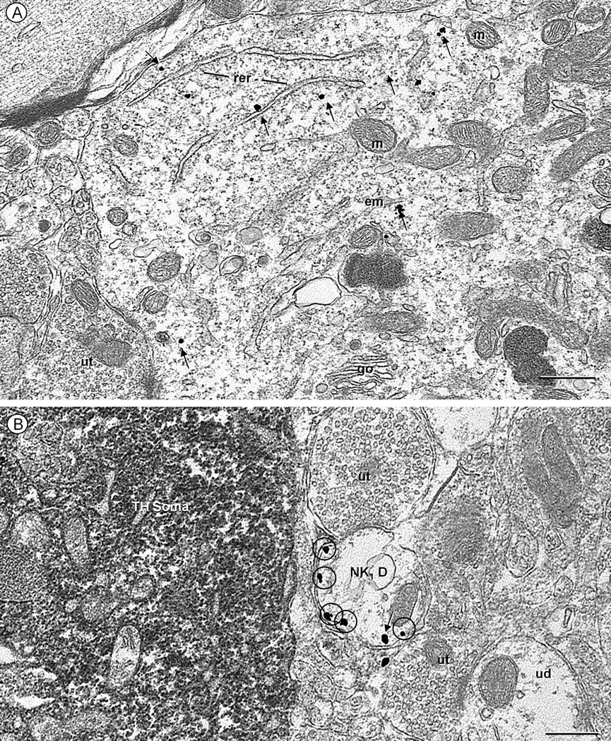
Fig. 2.
Electron micrographs displaying NK1 receptor (immunogold) in a soma (A) and dendrite (B) located within the cNTS of a control mouse. These structures are without detectable tyrosine hydroxylase (TH, immunoperoxidase) immunoreactivity, such as that seen in a nearby soma (TH-soma of B). A. NK1 immunogold particles (arrows) are seen throughout the cytosol in a soma receiving input from an unlabeled terminal (ut). The labeling is particularly evident near the rough endoplasmic reticulum (rer), Golgi apparatus (go) and endomembranes (em), but absent from mitochondria (m). B. Many extrasynaptic plasmalemmal NK1 immunogold particles (encircled) are seen in a transversely sectioned NK1-labeled dendrite (NK1 D). This dendrite is located in a neuropil that contains a TH-labeled soma as well as unlabeled terminals (ut) and dendrites (ud). Scale bar: 0.5 µm.

