Fig. 3.
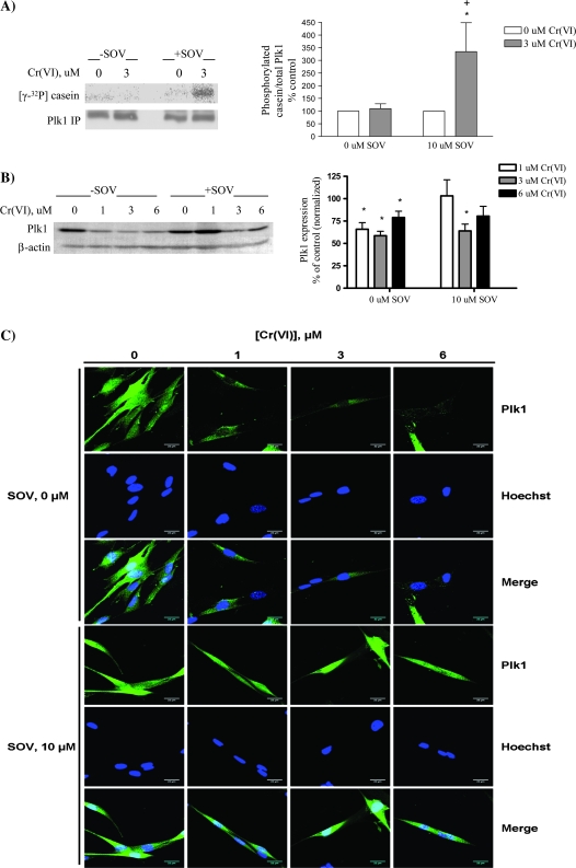
PTP inhibition during Cr(VI) exposure is associated with alterations in Plk1 activity, expression and localization. HLF cells were treated with the respective agents for 24 h, and cellular protein was extracted. (A) Plk1 was immunoprecipitated and used in an in vitro kinase assay with α-casein as a substrate. Proteins from each reaction were separated by sodium dodecyl sulfate–polyacrylamide gel electrophoresis, and Plk1 phosphorylation was detected by 32P incorporation. Plk1 protein in the immunoprecipitates was analyzed by western blotting. Data are expressed as percentage of control, in the absence of Cr(VI), and are the means ± standard error of four independent experiments. Asterisk indicates a statistically significant difference from the respective control at P < 0.05. Plus sign indicates a statistically significant difference between the samples treated with and without SOV at P < 0.05. (B) Total protein was separated by sodium dodecyl sulfate–polyacrylamide gel electrophoresis. Plk1 was detected by immunoblotting, and expression was normalized to that of β-actin. Data are expressed as percentage of control, in the absence of Cr(VI), and are the means ± standard error of four independent experiments. Asterisk indicates a statistically significant difference from the respective control at P < 0.05. (C) HLF cells were seeded in chamber slides and incubated with the indicated concentrations of Cr(VI) ± SOV. Cells were fixed and stained with Plk1 antibody followed by Alexa 488-conjugated fluorochrome. Cells were then stained with Hoechst nuclear dye and visualized at ×63 using a Zeiss laser scanning microscope with ZEN software, at excitation wavelengths of 488 and 405 nm for anti-Plk1 and Hoechst, respectively. Data represented in Results are the average of three independent experiments where at least 40 cells per experimental condition were scored per experiment. Images are representative of one independent experiment. Row 1: anti-Plk1 in the presence of Cr(VI); row 2: corresponding Hoechst staining of cells in row 1; row 3: merged images of row 1 and 2; row 4: anti-Plk1 in the presence of Cr(VI) and SOV; row 5: corresponding Hoechst staining of cells in row 4; row 6: merged images of rows 4 and 5.

