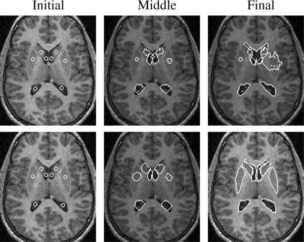
Fig. 7.

Segmentation of 8 sub-cortical structures (the lateral ventricles (λi = 0.8, ωi = 0.2), heads of the caudate nucleus (λi = 0.3, ωi = 0.7), and putamina (λi = 0.2, ωi = 0.8)) in a MR brain image without prior information (top) and with joint shape prior (bottom).
