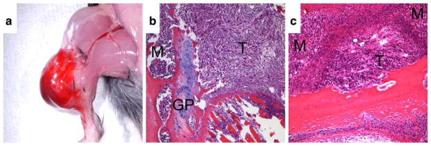
Fig. 2.
Gross and histological examination of RM1 injected tibia. (a) Gross analysis and (b) H&E stained section of a tibia 14 days following the implantation of 1 × 10⁴ RM1 cells. In (b), note that RM1 cells (T) have displaced the marrow of the tibial metaphysis and promoted extensive cortical lysis, subsequently invading adjacent soft tissue. Growth plate (GP) and marrow (M) of the epiphysis are indicated. Magnification of 10×. (c) H&E stained section of tibia 14 days following the injection of 1 × 10³ RM1 cells. A tumor nest (T) is shown adjacent to an area of focal cortical lysis and surrounded by marrow (M). Magnification of 20×

