Fig. 5.
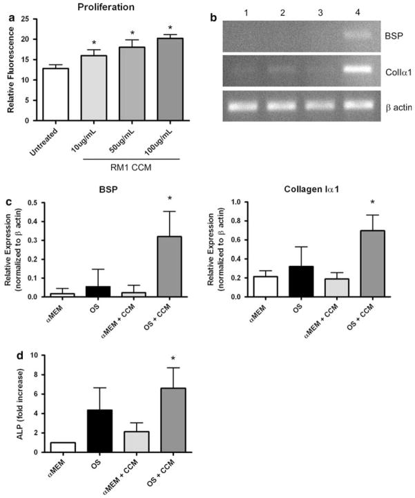
Effects of RM1 cells on osteoblast precursor cells in culture. (a) Osteoblast precursor MC3T3-E1 cells 3 days following treatment with increasing concentrations of RM1 conditioned culture media (CCM). (*) Treatments compared to untreated control (P ≤ 0.05). (b) Semi-quantitative RT-PCR of mRNA for genes involved in osteoblast differentiation from osteoblast precursor MC3T3-E1 cells. Results shown are representative of experimentation performed in triplicate. Treatments were as follows: (1) αMEM w/10% FBS, (2) OS medium (αMEM, 10%FBS, 50 μg ascorbate, 10 mM β-glycerol phosphate), (3) αMEM w/10% FBS and 50 μg/ml RM1 CCM, (4) OS medium with 50 μg/ml RM1 CCM. (c) Densitometric analysis of bands shown in (b). (*) OS + RM1 CCM compared to OS alone (P ≤ 0.05). (d) Quantification of surface ALP expressed by MC3T3-E1 cells 5 days after initiation of treatments. Treatments were as in (b) except for reduced serum supplementation (2% FBS). Bars represent area of ALP positive cells of 10 fields from experiments completed in triplicate. (*) OS + RM1 CCM compared to OS alone (P ≤ 0.05). All graphs are mean ± SD

