
Fig. 1.
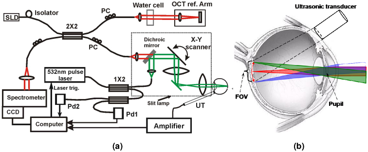
Experimental system of the OCT-guided PAOM. (a) Schematic of the OCT-guided PAOM. (b) Illustration of the optical beam delivery to the retina and the position of the ultrasonic transducer. The light delivery systems were built on a slit-lamp bio-microscope. The two imaging subsystems are synchronized by the PAOM laser pulses detected by photodiode Pd1. SLD: superluminescent diode; PC: polarization controller; Pd: photodiode; FOV: field of view; UT: ultrasonic transducer.