Figure 2.

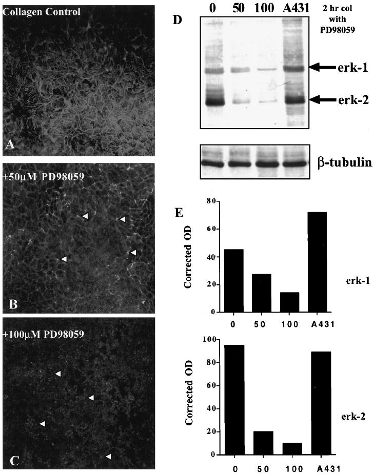
Epithelia treated with MAP kinase inhibitor, PD98059. Confocal images through the ACM optical plane (Fig. 1E, arrowhead) of whole epithelial tissue stained with FITC-phalloidin after incubation with PD98059 (50 and 100 μM; B, C) and stimulation with COL for 2 hours compared with control tissue (A). The lowest dose of PD98059 had little effect on ECM-stimulated ACM reformation; however, at the middle (B, arrowheads) and highest (C, arrowheads) doses, the blebs were more prominent. Western blot of activated erk-1 and -2 (D) showed a decrease in activity of these MAP kinase proteins from cell extracts that were treated with PD98059 in a dose-dependent manner compared with untreated epithelia (0) or control cell extracts (A431). The blot was reprobed with β-tubulin to demonstrate that the gel was loaded equally. Densitometry analyses (E) of the anti active erk-1 and -2 Western blot (D). β-Tubulin was used to obtain corrected densities (OD units) of the two proteins, erk-1 and -2. Both erk-1 and -2 decreased in a dose-dependent manner compared with uninhibited control tissue (0). Scale bar, 10 μm.
