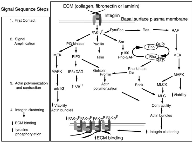
Figure 5.

The proposed signaling hypothesis. The stages of epithelial response to ECM were divided into four parts: 1) first contact, 2) signal amplification, 3) actin polymerization and contraction, and 4) integrin clustering leading to increased ECM binding and further signal amplification. Viewing the diagram from top to bottom, the first contact events occur at the basal cell surface but are drawn at the top of the diagram for illustration of the hypothesis. The first contact events include ECM binding and tyrosine phosphorylation of FAK. Signal amplification is represented by FAK phosphorylating the surrounding kinases and proteins (PI-3 kinase, paxillin, Fyn/ Shc, Src). These proteins amplify the signal further by stimulating their downstream targets leading to increased actin polymerization and contraction. The actin rearrangements aid increased integrin clustering and ECM binding producing a stronger intracellular signal through tyrosine phosphorylation.
