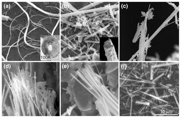
FIGURE 1.
Structure of asbestos fibers by transmission electron microscopy (TEM): (a) serpentine and (b–f) amphiboles. (a) International Union Against Cancer (UICC) asbestos chrysotile ‘A’ standard, (b) UICC asbestos crocidolite standard, Death Valley, California, (c) UICC asbestos anthophyllite standard, (d) winchite-richterite asbestos, Libby, Montana, (e) tremolite asbestos and (f) UICC asbestos amosite standard. Chrysolite is the only member of the serpentine group. Because of the mismatch in the spacing between the magnesium ions and the silica ions, chrysotile curls into a thin-rolled, flexible sheet while amphibole fibers are more rigid. Scale bar = 10 μm. (Reprinted with permission from Denver Microbeam Laboratory at the U.S. Geological Survey).

