
Figure 8.
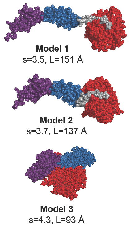
Models of the 61k fragment of smMLCK. Three different arrangements of the Ig2-Fn3 tandem with respect to the catalytic domain were tested. The domains are color coded: red - the catalytic/regulatory domain; blue - the Fn3 domain; magenta - the Ig(2) domain; gray - the linker region (linker-3) connecting the Fn3 domain with the catalytic domain. See Table 2 for the exact definition of the domains. Images of protein models were generated with the program PyMOL (85).