Figure 4.
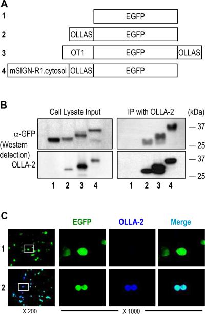
Immunodetection of OLLAS tagged EGFP proteins. (A) Schematic view of 4 recombinant EGFP proteins without a tag (#1) or with OLLAS epitope tag attached to the N-terminus (#2), C-terminus (#3), or internal site (#4). The OT1 peptide, a ligand of mouse MHC I from ovalbumin, were also present in the N-terminus of EGFP protein #3. The 53 amino acid full-length cytosolic domain from mouse SIGN-R1 was present at the N-terminus of EGFP protein #4. (B) Expression vectors for the 4 different recombinant EGFP proteins were transfected into 293T cells. Cell lysates (left) were immunoprecipitated (IP) with mAb OLLA-2 (right). Then, all samples were separated in SDS-PAGE gels followed by blotting with anti-GFP (upper panels) and mAb OLLA-2 (lower panels). (C) Expression vectors for EGFP (#1; upper panels) and EGFP with OLLAS tag at the N-terminus (#2; lower panels) were transfected into CHO cells. CHO cells were visualized with the signals for EGFP (green) and immunofluorescence staining for OLLA-2 (blue). Insets in left panels are shown at 1000 fold magnification in the right.

