Figure 5.
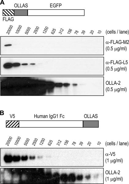
Comparison of binding sensitivity of mAb OLLA-2 and other anti-epitope tag mAbs. (A) 293T cells were transfected with pCMV-FLAG.OLLAS-EGFP (structure shown in upper panel), lysed, diluted as indicated, and separated in SDS-PAGE gels followed by blotting with 0.5 μg/ml of anti-FLAG mAb M2 (second panel), the new anti-FLAG mAb L5 (third panel), and mAb OLLA-2 (lower panel). (B) 293T cells were transfected with pCMV-V5-hIgG1Fc-OLLAS (structure shown in upper panel), lysed, diluted as indicated, and separated in SDS-PAGE gels followed by blotting with 1 μg/ml of anti-V5 mAb (Invitrogen; middle panel) and mAb OLLA-2 (lower panel).

