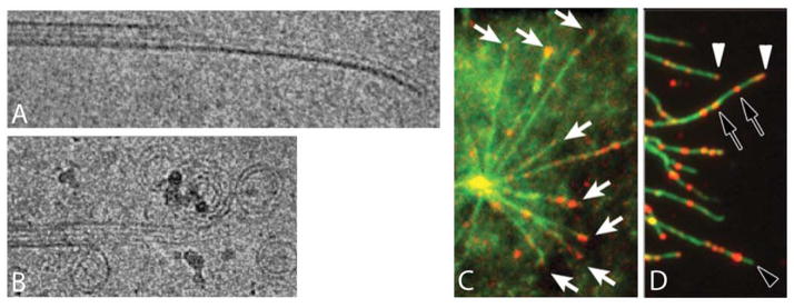
Figure 2.
Microtubule structural plasticity. A) Cryo-EM images of a growing microtubule end showing a curved, open sheet. B) Cryo-EM image of a shrinking microtubule end showing outwards curled protofilaments. C,D) Localization of microtubule segments with a stable lattice structure, recognized by a recombinant antibody. C. Pure tubulin microtubules (green) growing from a centrosome stained with an antibody that recognizes a stable structural state of the microtubule lattice (red). Note staining of growing tips (white arrows). D) Microtubules in a cell (green) stained with the antibody (red). Note tip staining, presumably on growing microtubles (white arrowhead), lack of tip staining, presumably on shrinking microtubules (empty arrowhead) and internal segments recognized by the antibody (empty arrows). A,B courtesy of T. Hyman, MPI Dresden. C, D adapted from (26).

