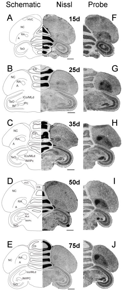
Fig. 11.

Developmental pattern of Cntnap2 expression in male posterior brain. Throughout development, Cntnap2 transcript distribution revealed a consistently robust hybridization signal in hemicoronal sections of male RA (F–J), as identified based on adjacent Nissl-stained sections (A–E). Strong Cntnap2 signals were also present in Cb, TeO and tectal nuclei IM/IPc (F–J). For abbreviations, see Table 1. Scale bars = 1 mm.
