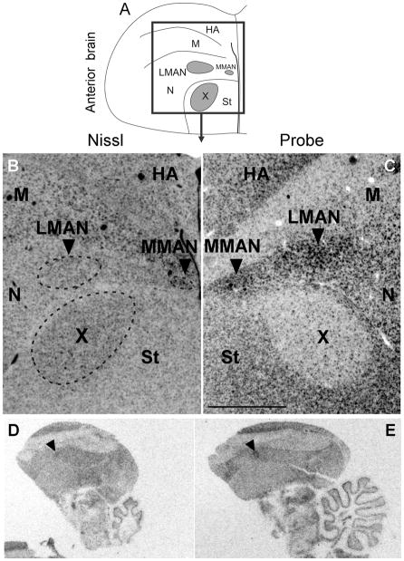
Fig. 5.
Inverted darkfield images of emulsion autoradiography of coronal sections of anterior adult male brain confirmed strong signals in LMAN and MMAN and low signals in area X compared to adjacent tissue (C). In situ hybridization of parasagittal sections of adult female brain indicate that there is considerable variability in Cntnap2 levels in LMAN, as identified by arrowheads in D and E. Unprocessed images in D and E, representing sections from different individuals, were taken under similar illumination conditions in order to emphasize the inter-individual variability in hybridization signals in female LMAN. For abbreviations see Table 1. Scale bar = 1 mm in C (applies to B and C).

