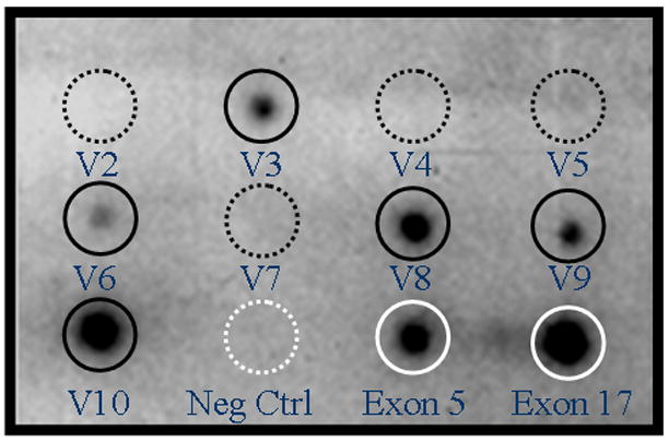
Figure 2.

Representative dot blot array hybridization of endometrial epithelial cells from a woman with endometriosis. Hybridization with specific splice variants showing v3, v6, v8, v9, and v10 (dark solid circles). Splice variants v2, v4, v5, and v7 are not expressed (dark dashed circles). Negative controls consisted of non-specific oligonucleotide sequences (white dashed circle). Positive controls are exon 5 and 17 from the non-variable region of CD44 (white solid circles).
