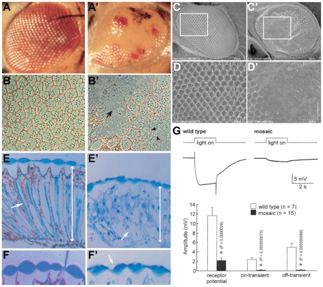
Figure 4. Large mosaic clones of Df(2R)Dg248 results in disruption of the adult eye.
Light and scanning EM micrographs of the wild type fly eye (A–F) show the classical ommatidial facets and bristles of the external eye. A′–F′, show the equivalent micrographs of fly eyes of Df(2R)Dg248 clones generated by the ey, FLP/FRT system. The Df(2R)Dg248 tissue in A′ is white while tissue with heterozygous expression of the wild type gene is red. The Df(2R)Dg248 (white) regions appear flattened and glossy (A′). Cross sections of the internal eye reveal that the normal (B) array of R-cells with central rhabdomeres is disrupted (B′ arrows) and there appears to be cell debris (B′ arrowhead) in these regions. Electron micrographs reveal regions where the external eye has collapsed and the facets have been obliterated (C′, D′). A transverse section through the eyes reveals the normal length (E, double arrow head) of the retina and its ordered array of ommatidia (arrow). The Df(2R)Dg248 (white) region appears disrupted (arrow) and the retinal thickness is shorter (double arrow head). The lenses, which normally (F) form a biconvex disc, are flattened on their external, but not internal, faces though the cuticle that forms the external boundary is visible within a flattened and partially “empty” lens (arrow, F′). (G) ERGs were recorded after a 5 min dark adaptation followed by a 2 sec bright-light pulses (top trace). The bottom trace shows representative ERGs of wild type (left) and Df(2R)Dg248 regions (white) of mosaic eyes. In Df(2R)Dg248 regions the 9-14 mV R cell depolarization (left) was greatly diminished and the early and late synaptic transients (left) completely abolished. Quantification of ERG parameters in wild type and Df(2R)Dg248 patches showed significant differences (P<0.01) between wild type and Df(2R)Dg248 eyes.

