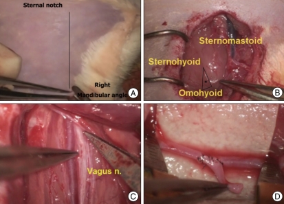
Fig. 4.
Microanastomosis training using the living rat model in the right cervical region. Skin is incised with scissor after hair removal (A). The triangle (*) limited by the sternomastoid, sternohyoid, and omohyoid muscles is located medial to the jugular vein (B). The common carotid artery and vagus nerve are exposed deep in this triangle (C). Trainee performs end-to-side anastomosis with an arterial graft (D).

