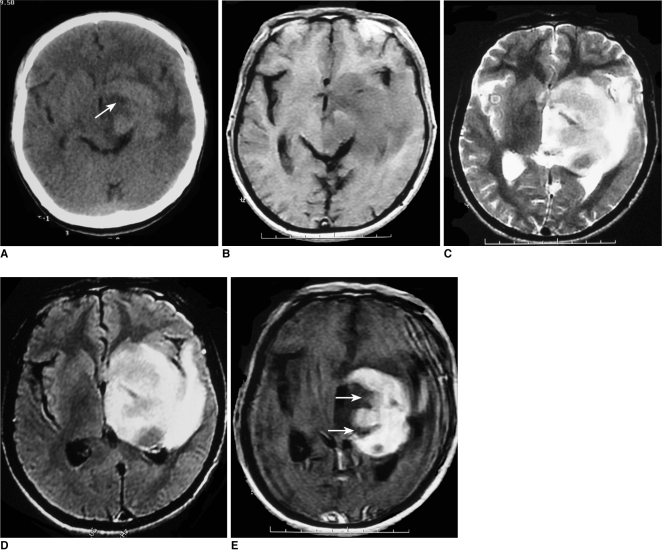
Fig. 1.
Images of 52-year-old man with lymphoma of left basal ganglia including plain CT image (A), axial T1-weighted image (B), T2-weighted image (C), FLAIR (D) and postcontrast axial T1-weighted image (E). Abnormally deep depression at tumor margin (notch sign) (arrow) was found on post-contrast axial T1-weighted image and plain CT image.

