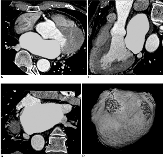
Fig. 2.
Measurement of left atrial volume using 3D volume threshold-based method of cardiac multidetector CT.
A-C. Axial, sagittal, and coronal views of left atrium. Endocardial contours of left atrium were traced on axial slices. Lowest value of CT attenuation was applied to cover contrast-enhanced whole left atrial cavity within region of interest. Included left atrial volume was confirmed by CT attenuation in three-orthogonal planes.
D. Volume-rendering threshold image of left atrium. Pulmonary vein confluences and atrial appendage were excluded from left atrial volume measurement.

