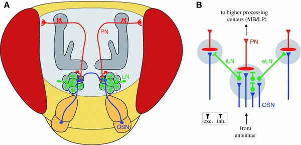
Fig. 2.

Schematic of the Drosophila olfactory pathway. a Antennal olfactory sensory neurons (OSN, blue) converge in specific glomeruli of the antennal lobe. Some of them send an axonal branch through the antennal commissure to the other hemisphere. Local interneurons (LN, green) branch in all glomeruli and interconnect these with each other. Projections neurons (PN, red) collect the olfactory information within the antennal lobe and send their axons to higher processing centers as the calyx and the lateral protocerebrum. b Circuit diagram of the antennal lobe. The three principal populations of neurons and their synaptic connections within the glomeruli (gray circles) are represented. The diagram summarizes anatomical data from several insect species. Excitatory synapses are symbolised by triangles, inhibitory synapses by bars. In Drosophila, the existence of both inhibitory local interneurons (iLN) as well as excitatory local interneurons (eLN) has been shown
