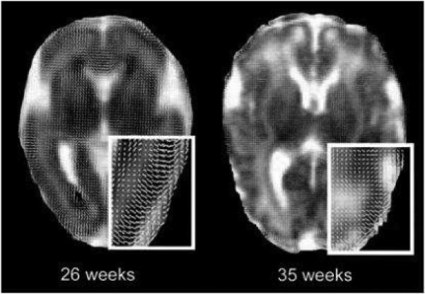
Fig 2.

Diffusion whisker plots overlaid on ADC images from infants of 26 and 35 weeks GA. The line segments are the projection of the major eigenvector onto the plane of the image and represent the orientation of the major eigenvectors. The insets with white borders are magnifications of the parieto-occipital regions of the images. Note that the major axes are oriented radially in cortex at 26 weeks GA. By 35 weeks GA, this feature is much less evident. In both images, organization of whiter matter is visible in the genu of the corpus callosum. The dark areas at the occipital horn of the lateral ventricles of the image from 26 weeks GA is due to small intraventricular hemorrhages layering dependently (with permission McKinstry et al. Oxford University Press)27.
