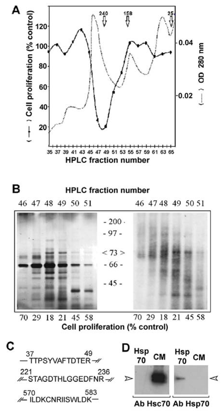
Figure 1. Identification of the 70 kDa-protein as the heat shock cognate protein Hsc70.

(A) Conditioned medium obtained from confluent 3Y1-Ad12 cells labelled with 35S-methionine was dialyzed, concentrated and purified on an HPLC system as described in the Materials and Methods. The UV elution profile was recorded at 280 nm (dashed lines). The antiproliferative activity of the collected fractions was measured by adding 75 μl of each odd fraction to triplicate culture wells containing 100,000 RBA cells growing in the log phase. The DNA content was evaluated after 5 days incubation and the values are expressed as a percentage of DNA recovered in control wells treated with buffer alone (solid lines). (B) HPLC fractions from the above experiment containing the antiproliferative activity were subjected to 12% SDS-PAGE separation without any reducing agent, and silver stained (left) or processed for fluorography of S35-labelled proteins (Brighty & Jassal). (C) Identification of rat Hsc70 by peptide microsequencing. The major 70–73 Kda protein was digested with trypsin as described in the Materials and Methods. Three different peptides were isolated by HPLC and sequenced. All sequences were identical to the rat 73 kDa heat shock cognate protein (Hsc70) and peptide 3 was specific to Hsc70 isoform 1. (D) Secreted proteins from conditioned medium (CM) or commercialized Hsp70 (Hsp70) were immunoblotted with Hsc70 or Hsp70 specific antibodies.
