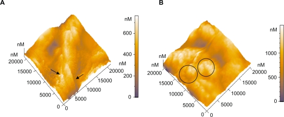
Figure 6.
Three-dimensional AFM topography image of: A) Platelets on type A carbon nanocoatings with 20% H2 in plasma during deposition after 1 hour incubation (scan size 21 μm × 21 μm). They form aggregations as presented with the arrows, with a mean maximum height of approximately 659 nm, B) Platelets on type A carbon nanocoatings with 20% H2 in plasma during deposition, after 2 hours of incubation (scan size 21 μm × 21 μm). The platelet clusters as denoted by the circles, have a height of more than 1000 nm.

