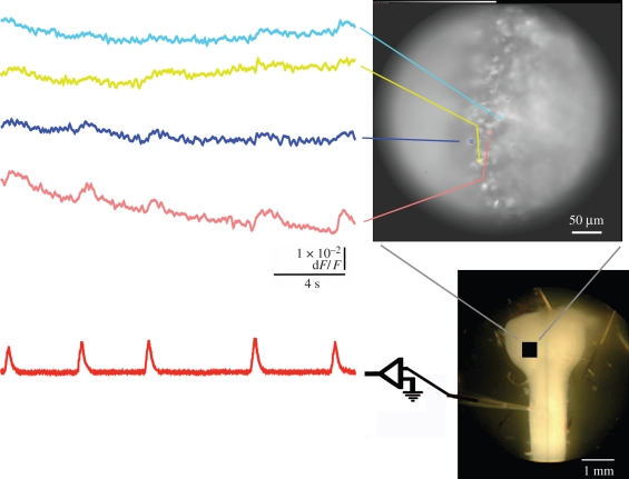
Figure 1.
Recordings of fluorescence changes (top left traces) in rostral medulla (right, top and bottom) and the simultaneous integrated activity recorded from the C4 ventral root (bottom left trace) in brainstem–spinal cord preparation of E18 mouse (5% CO2, pH 7.4) stained with Oregon Green BAPTA-1 AM, a calcium-sensitive dye, recorded optically and electrically, respectively, during a period of 20 s. Changes in fluorescence were averaged within coloured areas in the upper photograph and displayed as corresponding coloured traces on the left. A photograph of a similar preparation is shown (right, bottom) with a suction electrode attached to the ventral root and the approximate region of view indicated with a black square.

