Figure 5.
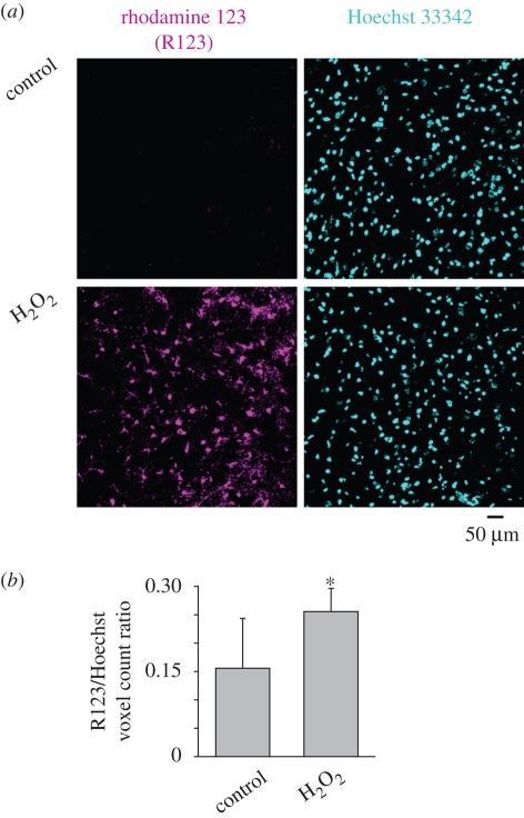
Oxidative stress of hypoglossal motoneurons induced by H2O2. (a) Histological example of rhodamine 123 staining of cells in hypoglossal slices incubated for 20 min in control solution or in the presence of H2O2 (1 mM). After rinsing, cells were stained with rhodamine 123 (5 µM; magenta pseudocolour) to reveal intracellular oxidative processes (Gomes et al. 2005; Jiang et al. 2006) and Hoechst 33342 (cyan pseudocolour) to show cell nuclei (Sharifullina & Nistri 2006). Note that, notwithstanding the analogous number of Hoechst-positive cells, there is a larger number of rhodamine 123-positive elements following H2O2. (b) Histograms show the ratio of rhodamine-positive cells over Hoechst-positive cells, indicating a significant rise after H2O2. Data are from 11 slices in control conditions and 14 slices after H2O2 and are quantified with ImageJ software (*p < 0.03). (F. Nani 2008, unpublished data).

