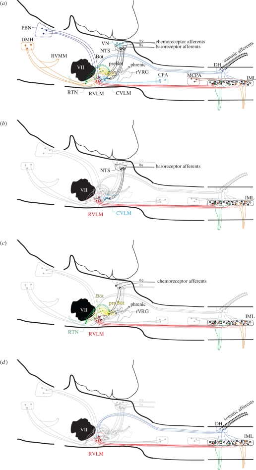
Figure 1.
A diagram of pathways in the regulation of the cardiorespiratory system: (a) all pathways overlapped. The bulbospinal red pathways are in the RVLM (figure 2a) and integrate information from the centre and the periphery. The output from this nucleus is crucial for maintaining normal sympathetic tone. PBN, parabrachial nucleus; DMH, dorsomedial hypothalamus; CVLM, caudal ventrolateral medulla; VLM, ventrolateral medulla; rVRG, rostral ventral respiratory group; CPA, caudal pressor area; MCPA, medullo cervical pressor area; IML, intermediolateral cell column; RVMM, rostral ventromedial medulla; VII, facial nucleus; RTN, retrotrapezoid nucleus; preBöt, preBötzinger neurons; VN, vestibular nucleus. (b) The baroreflex pathway is shown on its own. Stretch receptor afferent neurons from the aortic arch and carotid sinus and the neurons synapse in the nucleus tractus solitarius (NTS). Neurons in the NTS then activate inhibitory neurons (blue) in the caudal ventrolateral medulla, which in turn inhibit the neurons in the RVLM; this intense gamma-aminobutyric acid (GABA)-mediated inhibition inhibits sympathetic outflow, causing blood pressure and sympathetic nerve activity to fall. Note also the yellow respiratory neurons that modulate the activity of the cardiovascular neurons (also in c). (c) The pathways for peripheral and central chemoreceptors are shown. Central chemoreceptors are highly responsive to changes in CO2 and are found in the retrotrapezoid nucleus. Many of these chemosensitive neurons (greater than 40%) are galaninergic and Phox2b positive, but all lack tyrosine hydroxylase (Stornetta et al. 2009; figure 2b). Peripheral chemoreception emanates from the carotid body. Neurons terminate in the medial NTS (like the baroreceptors). From here, the excitatory information passes to both respiratory and cardiovascular neurons. (d) The somatosympathetic pathway is shown in an abbreviated form. Afferent nociceptive pathways enter the spinal cord in the dorsal roots, activate circuits locally, and at several stations throughout the neuraxis including the RVLM. This pathway is excitatory and results in the appearance of a variable number of peaks in sympathetic nerve activity, depending on which nerve is recorded from. In the case of the greater splanchnic nerve, this is generally two peaks.

