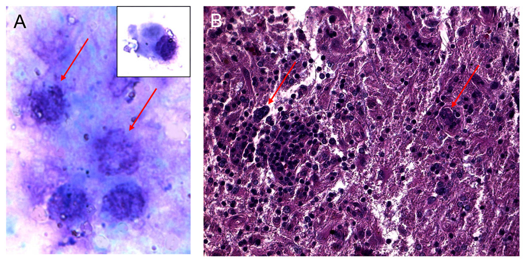
Figure 2.
A. Cytology of vitreous cells revealing reactive lymphocytes, plasma cell (insert), and large maligant lymphoma cells identifiable by their prominent nuclei, multiple nucleoli, and scanty basophilic cytoplasm. (Giemsa, original magnification, ×640)
B. Histology of brain biopsy showing large atypical lymphoid cells (arrows) infiltrated the CNS. (hematoxylin & eosin, original magnification, ×200)

