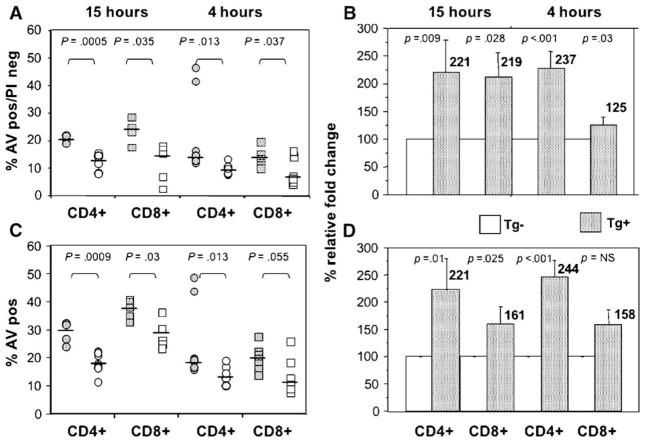
Fig. 2.
Increased apoptosis of activated CD4+ and CD8+ T cells after co-culture with transgenic hepatocytes expressing HCV core, E1 and E2 proteins. Apoptosis of activated T cells was studied by 3-color FACS analysis, 4 or 15 h after co-culture with HCV transgenic (Tg+, stippled) or non-transgenic (Tg−, open) hepatocytes. (A) T cells in the early phase of apoptosis (annexin V-positive, PI-negative) are shown. Increased apoptosis of activated T cells after co-culture with HCV transgenic (HCV Tg+) compared to non-transgenic (HCV Tg−) hepatocytes was seen at both 4 and 15 h and was seen for both CD4+ (circles) and CD8+ (squares) cells. Data of five to eight independent experiments were analyzed by Mann–Whitney U test. (B) The relative increase of early apoptotic cells increases after co-culture with Tg hepatocytes. In this example, data for Tg hepatocytes were normalized to the appropriate Tg− control. Results for both CD4+ and CD8+ remain statistically significant at 4 and 15 h. (C) All annexin V+ cells, showing increased apoptosis of both CD4+ and CD8+ T cells at 15 h, and an increase in CD4+ AV+ cells at 4 h. (D) Relative increase in all AV+ cells.

