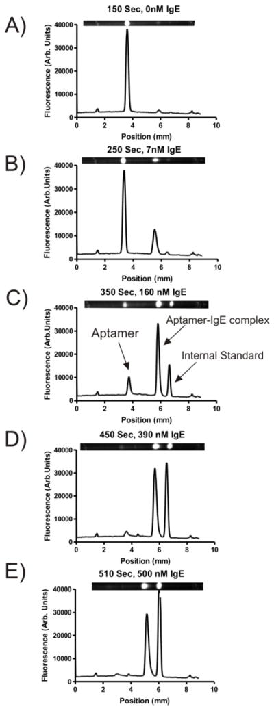
Figure 2.
Images and linescan plots recorded at various time points during an μFFE gradient where a constant concentration of fluorescently labeled aptamer for IgE was titrated with an increasing concentration of IgE. Peaks from left to right were identified as the unbound aptamer, the aptamer-IgE complex, and the internal standard (rhodamine 110). The anode is at the left side of the images.

