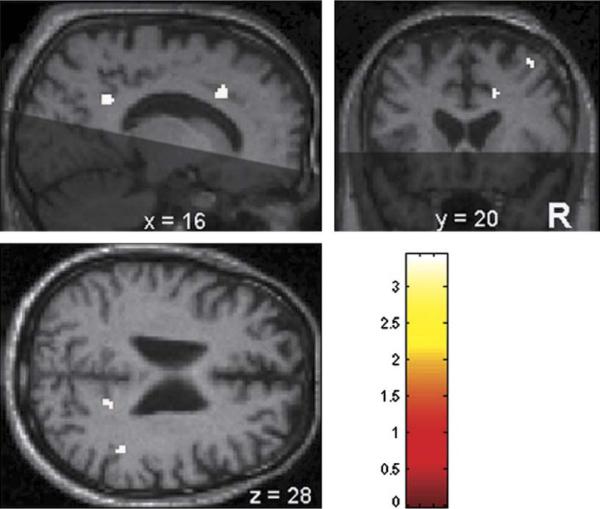
FIGURE 1.
Statistical parametric maps showing regions of hypoperfusion in the 13 mild cognitive impairment subjects who converted to dementia relative to the 35 mild cognitive impairment subjects who remain nondemented. Converters had hypoperfusion in the right precuneus/posterior cingulum (shown in the sagittal and axial slices), right middle cingulum (shown in the sagittal and coronal slices), right middle frontal cortex (shown in the coronal slice), and right inferior parietal cortex (shown in the axial slice). The shaded areas in the sagittal and coronal slices represent regions not covered by our implementation of arterial-spin labeling magnetic resonance imaging.

