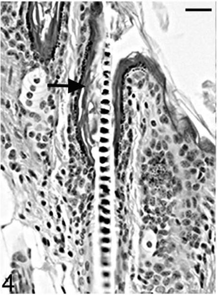
Figure 4.
Skin; Hrrh-R/Hrrh-R mouse, 14 days old. Mouse dorsal skin from the interscapular region was sectioned and stained with HE. Infundibula were mildly ectatic and filled with laminated cornified cells (black arrow). The stratum granulosum extended from the epidermis to the entrance of the sebaceous gland duct. Bar: 20 μm.

