FIGURE 1.
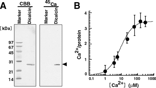
Ca2+ binding activity of Xenopus dicalcin. A, 45Ca blot of Xenopus dicalcin. Recombinant dicalcin and molecular size markers (∼6 μg each) were electrophoresed and transferred onto a PVDF membrane. Blots of recombinant Xenopus dicalcin and size markers were soaked in Tris-buffered saline containing 1 mm 45CaCl2. After washing, the membrane was dried, and bound 45Ca was detected. CBB, Coomassie brilliant blue staining; 45Ca, 45Ca blot; Marker, molecular size markers; Dicalcin, recombinant Xenopus dicalcin. B, Ca2+ binding to Xenopus dicalcin. Recombinant dicalcin (final concentration, 10 μm) was incubated with various concentrations of 45CaCl2. The graph shows the amount of Ca2+ bound to Xenopus dicalcin as a function of free Ca2+ concentration (n = 6, mean ± S.D.). The data were fitted by a Hill equation (solid line).

