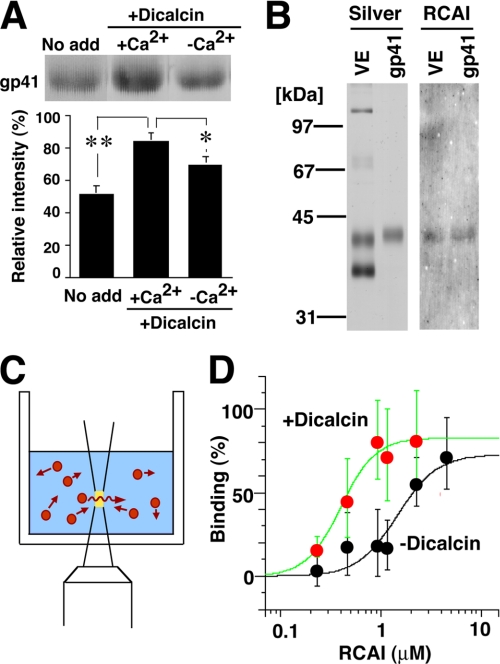
FIGURE 7.
Dicalcin binding to gp41 increases the reactivity of gp41 with RCAI. A, blots of VE proteins were preincubated in the presence or absence of dicalcin and Ca2+, followed by incubation with Rhodamine-labeled RCAI. Representative RCAI binding to gp41 was indicated (upper). The normalized intensity was significantly increased in the presence of dicalcin and Ca2+ (lower graph, n = 8; *, p = 0.06; **, p = 0.002). No add, nothing preincubated; +Dicacin, preincubation with dicalcin (1 μm); +Ca2+, preincubation in the presence of Ca2+ (500 μm CaCl2); −Ca2+, preincubation in the absence of Ca2+ (500 μm EGTA). B, isolation of gp41. Silver, silver-stained VE proteins and isolated gp41; RCAI, blot with RCAI. C, a scheme that represents FCS measurement. A fluorescent signal of diffusing molecule (red) was detected within a small defined confocal volume (yellow) of the well. Autocorrelation analyses of the fluctuating fluorescent signal estimate the diffusion time of fluorescent molecules and distinguish small fast diffusing (i.e. free fluorescent molecules) and large slow moving (target-bound molecules); this enabled us to investigate the stoichiometry of binding. D, the binding of TMR-labeled gp41 to RCAI was analyzed with FCS either in the presence of 1 μm dicalcin (red circles) or absence of dicalcin (black circles) (n = 15). Maximum and minimum diffusion time in each measurement was set to 100 and 0% binding, respectively. Each group of data were fitted with a Hill equation.

