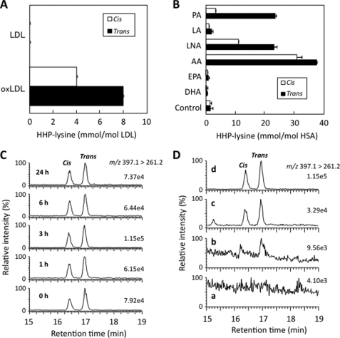
FIGURE 5.
In vivo formation of HHP-lysine via lipid peroxidation. A, LC/ESI/MS/MS analysis of cis- and trans-HHP-lysines in oxidized LDL (oxLDL). LDL (0.5 mg) was incubated with 5 μm Cu2+ in 1 ml of PBS at 37 °C. The native LDL and oxidized LDL were analyzed by LC/ESI/MS/MS with MRM mode followed by acid hydrolysis. B, LC/ESI/MS/MS analysis of cis- and trans-HHP-lysines in protein exposed to lipid peroxidation. The metal-catalyzed oxidation of unsaturated fatty acids in the presence of HSA was performed by incubating HSA (1 mg/ml) with 2 mm unsaturated fatty acids in the presence of 50 μm Fe2+ and 1 mm ascorbic acid in 1 ml of 50 mm sodium phosphate buffer, pH 7.2, in atmospheric oxygen at 37 °C. The native and modified HSA were analyzed by LC/ESI/MS/MS with MRM mode followed by acid hydrolysis. The abbreviations used are as follows: PA, palmitoleic acid; LA, linoleic acid; LNA, γ-linolenic acid; AA, arachidonic acid; EPA, eicosapentaenoic acid; DHA, docosahexaenoic acid. C, LC/ESI/MS/MS analysis of cis-[U-13C6,15N2]- and trans-[U-13C6,15N2]HHP-lysines in the kidneys of mice exposed to Fe3+-NTA. The ion current tracing of cis-[U-13C6,15N2]- and trans-[U-13C6,15N2]HHP-lysines LC/ESI/MS/MS with MRM is shown. D, LC/ESI/MS/MS analysis of cis- and trans-HHP-lysines in the kidneys of mice exposed to Fe3+-NTA. The ion current tracing of HHP-lysine using LC/ESI/MS/MS with MRM is shown. Panel a, 0 h after administration; panel b, 1 h after administration; panel c, 3 h after administration; panel d, internal standards.

