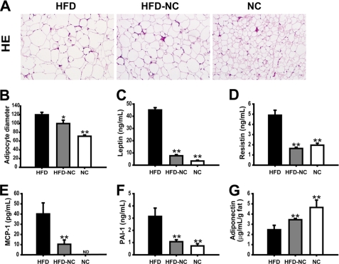
FIGURE 4.
Morphology of epi-WAT and plasma adipokine levels. Paraffin-imbedded epi-WAT sections were stained with hematoxylin and eosin (A), and the relative adipocyte diameter was measured by using Image J software (B). Multiple enzyme-linked immunosorbent assays were used to measure the concentration of leptin (C), resistin (D), MCP-1 (E), PAI-1 (F), and adiponectin (G) in serum of HFD, HFD → NC, and NC mice. Data are expressed as the mean ± S.E., n = 9–10 per group in B–G and J. **, p < 0.01 compared with the HFD group. ND means not detectable.

