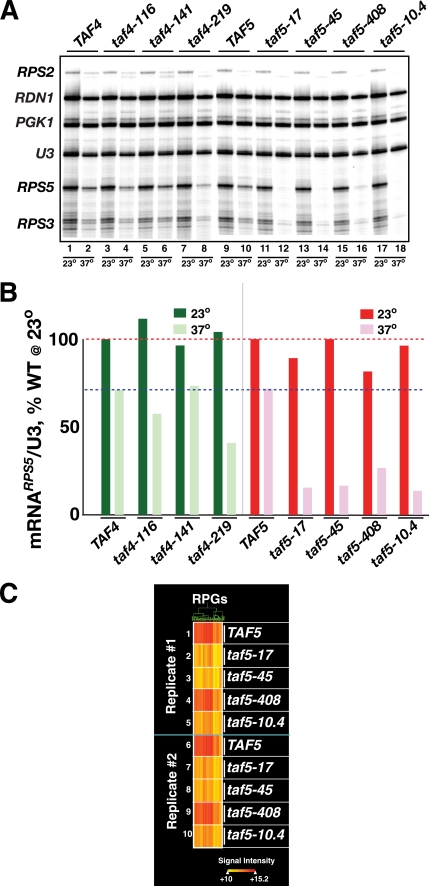
FIGURE 3.
Effect of temperature shift of taf4 Ts+ and taf5 Ts+ strains on RPG transcription. A, log phase cells expressing the indicated TAF4 and TAF5 alleles (top) were grown at 23 or 37 °C for 2 h and harvested, and total RNA was extracted. Equal amounts of RNA were subjected to multiplex primer extension analyses using a mixture of 5′-32P-labeled gene-specific oligonucleotide primers (RPS2, RDN1, PGK1, U3, RPS5, and RPS3). Extension products were fractionated on a sequencing gel that was dried, exposed to a Kodak K-screen, and scanned with a Bio-Rad FX imager; relevant portion of image is shown. B, scan was analyzed using Bio-Rad QuantityOne software. RPS5 mRNA-specific signals were normalized to WT and U3 signals and plotted; dashed lines represent signals of WT at 23 °C (top line, red) and 37 °C (bottom line, blue). C, effect of taf5 mutations on expression of the RPG regulon. The 37 °C TAF5 and taf5 Ts+ total RNA samples analyzed in A and B above (Replicate #1, 1–5), as well as the equivalent RNA from an independent biological replicate (Replicate #2, 6–10) were Cy5-labeled and hybridized to Nimblegen oligonucleotide yeast genome arrays. The hybridization signals of the RPGs whose expression changed by 2-fold (up- or down) in the taf5-17-derived sample relative to TAF5 are plotted in heat map format following hierarchical clustering. Hybridization signal intensity is indicated by the “heat” scale shown (bottom).

