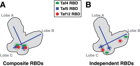
FIGURE 7.
Possible organization of Taf4, -5, and -12 RBDs within the TFIID complex. Shown is a schematic of yeast TFIID with A, B, and C lobes labeled (13). TFIID contains two copies of both Taf5 and the Taf4/Taf12 heterodimer (5), and all three subunits have been immunomapped within the structure. The N termini of the two molecules of Taf5 map to lobe C, whereas the two Taf5 C termini map to lobes A and B. The localization and orientation of the Taf5 subunits are indicated by the blue lines, whereas Taf4, -5, and -12 RBDs are indicated by the green ovals, blue squares, and red circles, respectively. A, model depicting two tightly localized Rap1-binding sites (RBDs, two dashed circles) composed of combined Taf4, -5, and -12; Composite RBDs. B, model depicting six independent Rap1-binding sites within Taf4, -5, and -12 (six dashed circles); Independent RBDs. Note that the currently available resolution of S. cerevisiae TFIID subunits is too low to assign precise locations for the Taf5 N termini and Taf4/12 heterodimer pairs (17).

