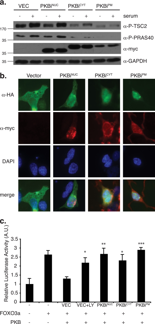
FIGURE 2.
Subcellular compartment-restricted PKB/Akt inhibition. a, NIH 3T3 cells transduced with retrovirus encoding localized myc-tagged PKBis were starved in medium containing 0.2% FBS overnight. Starved cells were either left untreated or stimulated with 10% FBS for 15 min. Total cell lysates were normalized for protein content and 25 μg was separated by SDS-PAGE followed by transfer to PVDF membrane and immunoblotting with the indicated antibodies against cytoplasmic PKB/Akt substrates (P-TSC2, P-PRAS40), epitope-tagged inhibitors (myc), and loading control (GAPDH, glyceraldehyde-3-phosphate dehydrogenase). b, 3T3L1 cells were transiently co-transfected with cDNA encoding HA-tagged FOXO3a and myc-tagged localized PKBis. Co-staining was performed using fluorescein-conjugated α-HA antibody (green) and indirect immunofluorescence against the myc tag (red). c, COS7 cells were transiently co-transfected with the pGL3-FOXO luciferase reporter of FOXO activity, FOXO3a, PKB/Akt, the indicated localized PKBi constructs, and equal amounts of β-galactosidase cDNA. LY294002 (LY) treatment was 12.5 μm overnight. Luciferase activity was assayed and normalized to β-galactosidase activity to account for differences in transfection efficiency. Three experiments were performed in triplicate and values were plotted relative to basal pGL3-FOXO luciferase activity (error bars = S.E.; *, p < 0.02; **, p < 0.01; ***, p < 0.001). DAPI, 4′,6-diamidino-2-phenylindole.

