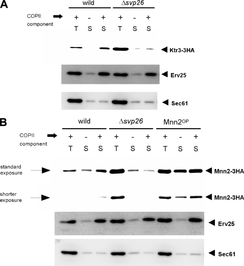
FIGURE 4.
In vitro COPII budding assay using either wild-type or Δsvp26 membranes. A, the ER-enriched membrane fractions prepared from the indicated strains were incubated either in the presence (+) or absence (-) of purified COPII coat components and the incorporation of Ktr3-3HA into COPII vesicles was analyzed by immunoblotting. T contains 2.5% of the total reaction mixture, and S contains 75% of the total COPII vesicle fraction, both of which were collected after the reaction. Erv25 and Sec61 were monitored as a positive and negative control of the experiment, respectively. B, incorporation of Mnn2-3HA into COPII vesicles was analyzed as in A. An assay using microsomal membranes prepared from the Mnn2OP cells were also performed. For detection of Mnn2-3HA, in addition to the standard exposure, a shorter exposure of the same blot is also shown to indicate the differences in intensity more clearly.

