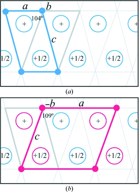
Figure 4.
Selected indexed unit cells. Simplified representation of packing diagrams for the (a) small and (b) doubled monoclinic cells. Open circles represent HCA II molecules. In (a) the blue cell outlined is the selected indexed small monoclinic cell (a ≃ 42, b ≃ 41, c ≃ 73 Å, β ≃ 104°) and contains one molecule (blue open circle) in the asymmetric unit. The gray cell outlined is an alternative but not selected small monoclinic cell (a ≃ 42, b ≃ 41, c ≃ 74 Å, β ≃ 109°) that was not selected in indexing as the c axis is larger. In (b) the red cell outlined is the selected indexed doubled monoclinic cell (a ≃ 84, b ≃ 41, c ≃ 74 Å, β ≃ 109°). This is the a-doubled cell of the not-selected small monoclinic cell shown in (a) and contains two molecules in the asymmetric unit. The blue open circles are the A-chain ordered molecules and the red open circles are the rotationally disordered B-chain (B′ and B′′) molecules. This figure was produced with PyMOL (DeLano, 2002 ▶).

