Figure 3.
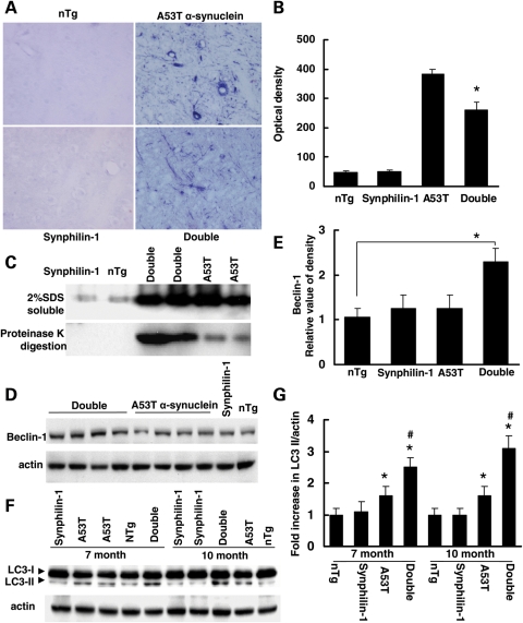
Abnormal accumulation of α-synuclein was less in double-transgenic mice at the preclinical stage. (A and B) Brain sections from mice at 7 months of age (preclinical stage) were digested with proteinase K for 3 h and then followed by immunostaining using anti-α-synuclein antibodies. Double-transgenic mice displayed less accumulation of abnormal α-synulcein after proteinase K digestion compared with A53T mice at 7 months of age. (A) Representative images of anti-α-synuclein immunostaining of various experimental groups after proteinase K digestion. (B) Graph showing the quantification of α-synuclein accumulation after proteinase K digestion in pons at 7 months of age. (C) Western blot analysis of brainstem homogenates of mice at 7 months of age (preclinical stage) using anti-α-synuclein antibodies. Top, 2% SDS-soluble homogenates; bottom, homogenates were digested with proteinase K followed by western blot analysis using anti-α-synuclein antibodies. (D) Western blot analysis of brain homogenates of mice at 7 months of age (preclinical stage) using anti-beclin-1 antibodies. (E) Graph showing the quantification of (D). P < 0.05 by ANOVA. (F) Western blot analysis of brainstem homogenates (2% SDS-soluble samples) of mice using anti-LC3 and anti-actin antibodies. (G) Graph showing the quantification of (F). *P < 0.05 by ANOVA versus aged-matched non-transgenic control mice or synphilin-1 transgenic mice; #P < 0.05 by ANOVA versus aged-matched A53T single-transgenic mice.

