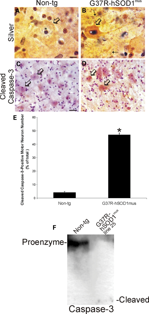
Figure 10.
Degeneration of spinal MNs in hSOD1mus tg mice is apoptotic-like. (A and B) Silver staining in control mouse spinal cord showed that MN cell bodies (A, open arrow), their nuclei (A, asterisk) and the surrounding neuropil were free of degeneration, whereas mutant-hSOD1mus tg mouse (line 73) MNs showed somatodendritic attrition with nuclear condensation (B, open arrow), and the neuropil contained numerous degenerating processes (B, hatched arrows). Scale bar (A and B): 8 μm. (C and D) In spinal cord sections stained immunohistochemically for cleaved capsase-3 (using immunoperoxidase with DAB, brown staining), faint granule-like labeling was observed in the cytoplasm of few MN cell bodies in 12–15-month-old controls (C), whereas age-matched mutant-hSOD1mus tg mice (line 73) showed accumulation of cleaved caspase-3 in the cell body of subsets of MNs (D, open arrows). Sections were counterstained with CV. Scale bar (C and D): 28 μm. (E) Graph of the number of lumbar MNs that were positive for cleaved caspase-3 in control non-tg mice and hSOD1mus tg mice expressing mutant hSOD1. Values are mean ± SEM (different lines for each construct are grouped). Mutant-hSOD1mus tg mice had a significant increase (P < 0.01) in the number of cleaved caspase-3+ MNs. (F) Western blot for caspase-3 in soluble protein extracts of total spinal cord of a mutant-hSOD1mus tg mouse (line 25 is shown) and an age-matched non-tg mouse. In non-tg mouse spinal cord, full-length 32 kDa pro-enzyme is detected strongly, but no cleaved (12 kDa) subunit is detected. In contrast, in G37R-hSOD1mus tg mouse spinal cord, the pro-enzyme level is reduced markedly and cleaved subunit is detected.

