Figure 2.
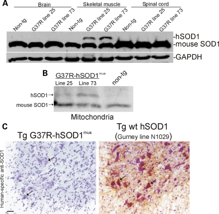
hSOD1mus tg mice express hSOD1 in skeletal muscle but not in CNS. (A) Soluble proteins were extracted from hind-leg skeletal muscle, spinal cord and brain of hSOD1mus tg (different mouse lines are identified) and non-tg mice. Samples (100 μg of protein loaded/lane) were electrophoresed in 16% SDS–PAGE gels, electroblotted onto nitrocellulose membrane and immunoblotted using an antibody that binds an epitope common to hSOD1 and mouse SOD1. The blot was re-probed with an antibody to GAPDH as a protein-loading control. (B) Proteins from mitochondria-enriched fractions were extracted from hind-leg skeletal muscle (gluteus, biceps and gastrocnemius pooled) and subjected (250 μg of protein) to immunoprecipitation for SOD1 and then western blotting for SOD1. hSOD1 is detected in mitochondria-enriched fractions of hSOD1mus tg mice (two different lines of G37R-hSOD1mus tg mice are shown) but not in non-tg mice. Endogenous (mouse) SOD1 is detected in mitochondria of hSOD1mus tg and non-tg mice. (C) Immunohistochemical confirmation that hSOD1 is not present in spinal cord and specifically is not in MNs (arrows) of hSOD1mus tg mice (line 25 is shown). Spinal cord sections were reacted with a mouse monoclonal antibody specific for hSOD1, and antibody binding was detected using immunoperoxidase with DAB as chromagen (brown reaction product) and counterstained with CV. Spinal cord sections from tg mice (line N1029, ref. 45) expressing human wild-type SOD1 ubiquitously in a tissue non-specific pattern was a positive control. All spinal MNs in hSOD1mus tg mice are negative (C, left hatched arrows). Most MNs in N1029 mice are strongly positive for hSOD1 (C, right, hatched arrows). Scale bar: 35 μm.

