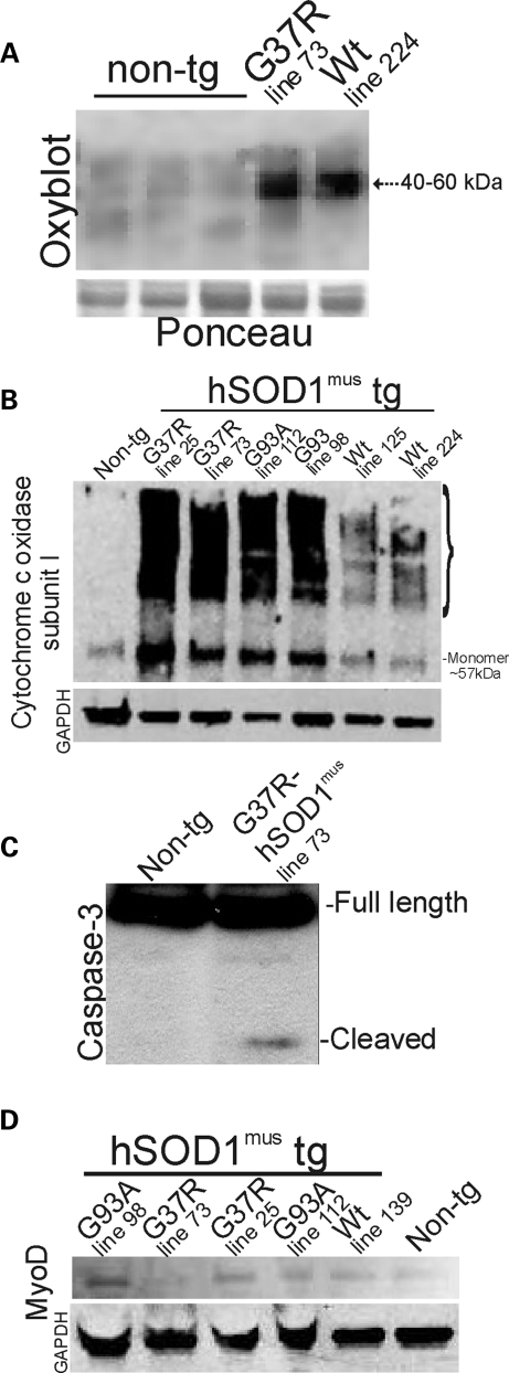
Figure 5.
Immunoblots confirm histological evidence of degenerative changes in skeletal muscle of hSOD1mus tg mice and show evidence for regenerative changes. (A) OxyBlot for carbonyl-modified proteins in the mitochondria-enriched fraction of 2–4-month-old non-tg and hSOD1mus tg mice. Tg mice expressing mutant and wild-type (Wt) variants of hSOD1, lines 73 and 224, respectively, have increased oxidative damage to proteins compared with controls. The same blot was stained with Ponceau S to show protein loading. (B) Western blot for Cox-I in the mitochondria-enriched fraction (5 μg of protein per lane) of 2–4-month-old non-tg and hSOD1mus tg mice. In non-tg mice, Cox-I is detected primarily as a monomer at ∼57 kDa (far left lane). The levels are elevated, and numerous higher molecular weight forms of Cox-I are formed in hSOD1mus tg mice expressing G37R mutant (lines 25 and 73) and G93A mutant (lines 112 and 98). hSOD1mus tg mice expressing wild-type hSOD1 (lines 125 and 224) also showed higher molecular weight species of Cox-I. The blot was re-probed for GAPDH as a protein-loading control. One G93A lane was underloaded. (C) Western blot for caspase-3 in the soluble protein fraction (100 μg of protein per lane) of skeletal muscle of 2–4-month-old non-tg and hSOD1mus tg mice. Cleaved subunit was detected in G37R-hSOD1mus tg mice (line 73). Full-length pro-enzyme, but not the cleaved subunit, was detected in age-matched non-tg mice. (D) Western blot for MyoD in the nuclear-enriched fraction (50 μg of protein per lane) of 2–4-month-old non-tg and hSOD1mus tg mice. MyoD levels are elevated in G37R mutants (lines 73 and 25), G93A mutants (lines 98 and 112) and wild-type (Wt, line 139) hSOD1mus tg mice compared with non-tg mice. The blot was re-probed for GAPDH as a protein-loading control.

