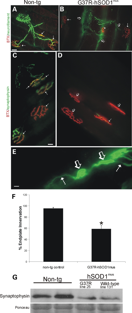
Figure 6.
hSOD1mus tg mice develop NMJ abnormalities, including loss of endplate occupancy, axonopathy and loss of pre-synaptic terminals. (A and B) Hind-leg skeletal muscle (gluteus, biceps femoris and medial gastrocnemius muscles) was dual-labeled with antibody to neurofilament (green) and BTX-conjugated Texas Red to identify MN axon innervation of motor endplates in age-matched non-tg mice and hSOD1mus tg mice. Control mice had normal appearing MN distal axons with uniform morphology and nearly complete occupancy of motor endplates with MN axons (A, hatched arrows). By 12 months of age, mutant hSOD1mus tg mice (line 25 is shown) had highly irregular shaped (dystrophic) MN distal axons (B, arrow), some of which were swollen and with apparent sprouts, and motor endplates were denervated (B, open arrows). Some (∼33%) of the motor endplates in hSOD1mus tg mice appeared small and collapsed (B, asterisk). Scale bar (A and B): 25 μm. (C and D) Hind-leg skeletal muscle (gluteus, biceps femoris and medial gastrocnemius muscles) was dual-labeled with antibody to synaptophysin (green) and BTX-Texas Red to identify MN pre-synaptic bouton innervation of motor endplates in age-matched non-tg mice and hSOD1mus tg mice. Control mice had richly ramifying pre-synaptic axon terminals with precise, near-complete innervation of the motor endplates (C, hatched arrows, yellow). In mutant hSOD1mus tg mice (line 25 is shown), many motor endplates lacked pre-synaptic innervation (D, open arrows) or were only sparsely innervated by MN axon boutons (D, hatched arrow). Scale bar (C and D): 20 μm. (E) Intramuscular axon in G37R- hSOD1mus tg mouse (line 25) showing axonopathy as seen by swollen and tortuous segments (open arrow) interspersed between normal segments of axon (hatched arrows). Scale bar: 10 μm. (F) Graph showing counts of the number of motor axon-innervated endplates in medial gastrocnemius muscle of mutant hSOD1mus tg mice (all G37R lines are grouped). Values are mean ± SEM, n = 3–6 mice per group. Asterisk denotes significant difference (P < 0.05) from control. (G) Western blot for the pre-synaptic vesicle protein synaptophysin in skeletal muscle extracts of age-matched (12–15 months) non-tg mice and hSOD1mus tg mice expressing G37R (line 25) or wild-type (line 131) variants. Equivalent protein loading is shown by the Ponceau S-stained membrane.

