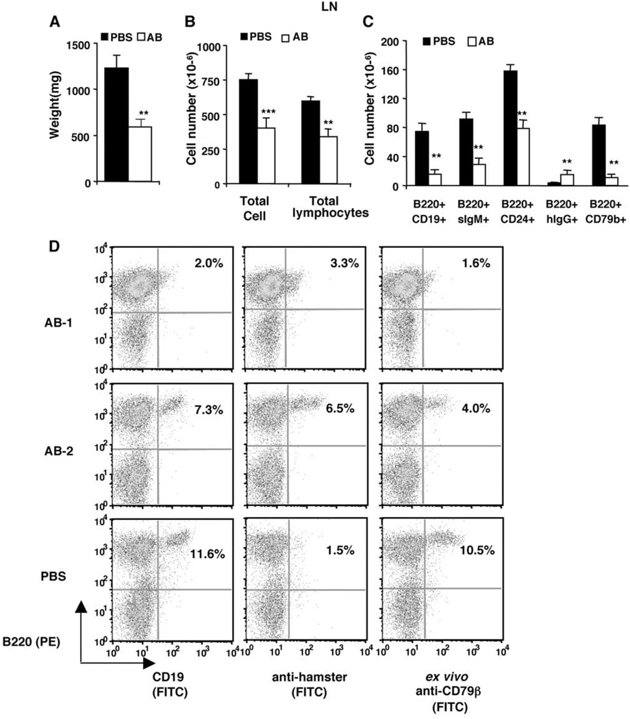
FIGURE 4.
B cell depletion from lymph nodes. Data from the same experiment as described in Fig. 3. A–C, Means and SEM of weight, lymphocyte numbers, and B cell numbers of lymph nodes. D, Representative anti-hamster IgG staining and binding to ex vivo Anti-CD79β by lymph node cells. Significant differences (*, p < 0.05; **, p < 0.01; ***, p < 0.001) were between AB and control-treated group.

