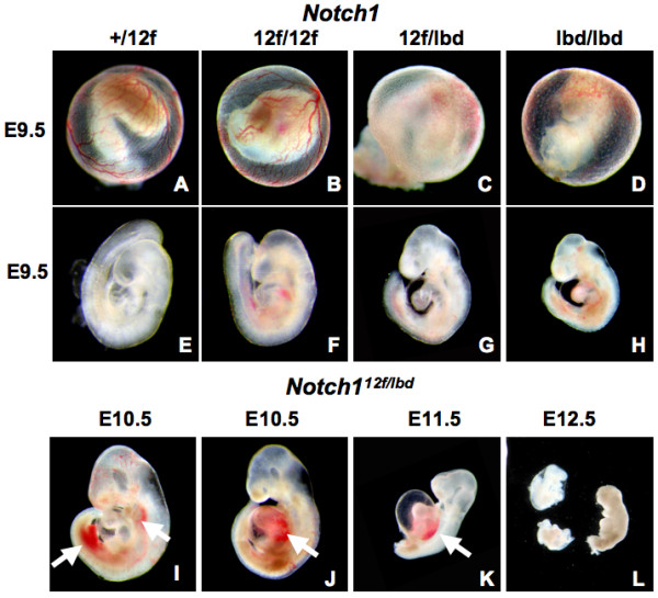
Figure 3.

Embryogenesis in Notch112f/lbd embryos. (A-D) Vascularization of yolk sac in Notch1+/12f, Notch112f/12f, Notch112f/lbd and Notchllbd/lbd embryos at E9.5. Large vitelline blood vessels were present in Notch1+/12f and Notch112f/12f yolk sacs, but absent in the Notch112f/lbd and Notch1lbd/lbd mutants. (E-H) Morphology of embryos at E9.5. Notch112f/12f are similiar to Notch1+/12f, Notch112f/lbd are markedly underdeveloped, and Notch1lbd/lbd are severely underdeveloped. (I-L) Notch112f/lbd embryos from E10.5-E12.5. White arrows show hemorrhaging in E10.5 and E11.5 embryos; most E12.5 embryos were resorbing. The number of embryos examined at each stage is given in Table 1.
