Figure 6.

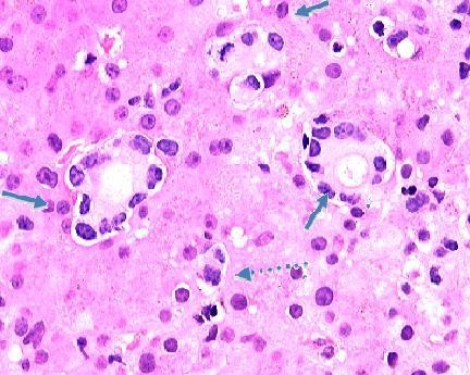
630× magnification of photomicrograph showing metastatic breast carcinoma cells infiltrating the liver sinusoids. Note the duct-like structures (arrows) with lining cells that have lost nuclear polarity and the mitotic form (dashed arrow).

630× magnification of photomicrograph showing metastatic breast carcinoma cells infiltrating the liver sinusoids. Note the duct-like structures (arrows) with lining cells that have lost nuclear polarity and the mitotic form (dashed arrow).