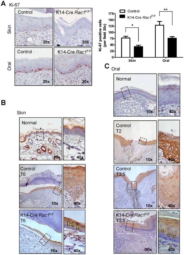
Figure 6. Rac1 excision leads to decreased cell proliferation and limited expression of cytokeratin 6, a stress and growth-related marker, in the skin and oral mucosa after wounding.
(A) Impairment of epithelial cell proliferation in the wounded area of the skin and oral mucosa of K14Cre Rac1F/F mice. Sections of epidermis adjacent to wounds healing skin (6 days after injury) and oral mucosa wounds (3.5 days after wounding) from of K14Cre Rac1F/F and control mice were subjected to Ki-67 staining as a marker of cell proliferation. Both anatomical sites of the K14Cre Rac1F/F mice show clear reduction in the proliferation capacity. Bar chart represents the total number of Ki-67 positive cells in the skin and oral cavity of K14Cre Rac1F/F and control mice (**p<0.01, * p<0.05). (B) Normal interfollicular skin does not express detectable levels of cytokeratin 6 (K6). The epithelial tongue adjacent to the wound area from normal skin (control) show remarkable up regulation of K6 in all the epithelial layers after 6 days of injury. K14Cre Rac1F/F mice fail to express K6 in the basal layer of the epithelial tongue from the skin (yellow arrows in the high magnification insert). (C) Normal oral mucosa does not express K6 during homeostasis (Normal). Expression of K6 in the oral mucosa epithelial tongue of control mice is evident at day 2 and 3.5 after injury. Expression pattern of K6 in the oral mucosa wound healing follows the skin pattern. K14Cre Rac1F/F mice show lack of K6 staining in the basal layer of the oral mucosa adjacent to the wound site as depicted (yellow arrowhead).

